
CME INDIA Case Presentation by Dr. D.P. Khaitan, Senior Consultant in Medicine, Ahmedabad; Dr. B.K. Shukla, Senior Consultant in Medicine, Arrah (Bihar).
CME INDIA Case Study
Abstract
Background: Tetralogy of Fallot with pulmonary hyperemia refers to a specific condition wherein the sufferer with this congential cyanotic heart disease experiences an increased blood flow to the lungs leading to pulmonary hyperemia. This is considered as atypical presentation of this condition. Most of cases of cyanotic TOF have oligaemic lung field due to Right ventricular outlet obstruction, or pulmonary stenosis in association with Right-to-left shunting of blood resulting in poor perfusion of lung. On Chest X-Ray P-A view this exhibits an oligaemic and mildly hyerlucent lung field. But a few 2 to 5 % cases show increased vascular markings on chest radiology due to accompanying pulmonary hyperemia.
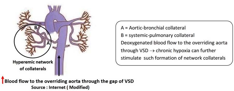
Case presentation: 4 yrs male child on 2D cardiac echo shows the evidence of Tetralogy of Fallot with perimembranous moderate to large sized VSD with narrow RV outlet and moderate post-valvular main PA stenosis, dilated RV (RVH), with aortic override (Less than 50%). And chest X-ray PA-view exhibits crisscross pulmonary hyperemia, possibly resulting from increased blood flow through aortic-bronchial and systemic- pulmonary collaterals.
Conclusion: The main aim of this case study (TOF) is to discuss the mechanism of pulmonary hyperemia in association with this condition and the way how to recognise this.
Introduction
Tetralogy of Fallot (TOF) is a complex congenital heart defect characterized by four anatomical abnormalities: ventricular septal defect (VSD), pulmonary stenosis, right ventricular hypertrophy, and an overriding aorta. These anatomical defects disrupt normal blood flow and oxygenation, leading to cyanosis and associated other clinical manifestations. TOF is one of the most common cyanotic congenital heart conditions, accounting for approximately 10% of all congenital heart disease. Most cases of cyanotic TOF have oligaemic lung field due to right ventricular outflow obstruction or pulmonary stenosis in association with right-to-left shunting of blood leading to poor pulmonary perfusion, witnessed on chest X-Ray PA view as an oligaemic and mildly hyperlucent lung field but 2 to 5 % of cases with TOF exhibit pulmonary hyperemia. This crisscross pulmonary hyperemia represents chronic volume overload, being transmitted to the lungs through the network of collaterals.
How Presented?
4 yrs male child, a suspected case of cyanotic congential heart defect was investigated with the following search out:
2D cardiac echo:
CYANOTIC CHD
VSD PERIMEMBRANOUS MOD TO LARGE SIZE
BIDIRECTIONAL FLOW
PREDOMINANT RT TO LT SHUNT 80% AND LT TO RT SHUTNT 20% (VISUAL) /
NON-RESTRICTIVE FLOW
PEAK VSD GR ——–12.5 mm Hg (RT TO LT) & 4 mm Hg (LT TO RT)
VSD SIZE ——0.9 CM
NARROW RV OUTLET AND MOD. POST VALVULAR MAIN PA STENOSIS
DILATED RV RA RVH
AORTIC OVERRIDE (LESS THAN 50%)
PASP 18 mm Hg
IMP —- CYANOTIC CHD – TETRALOGY OF FALLOT
ECG
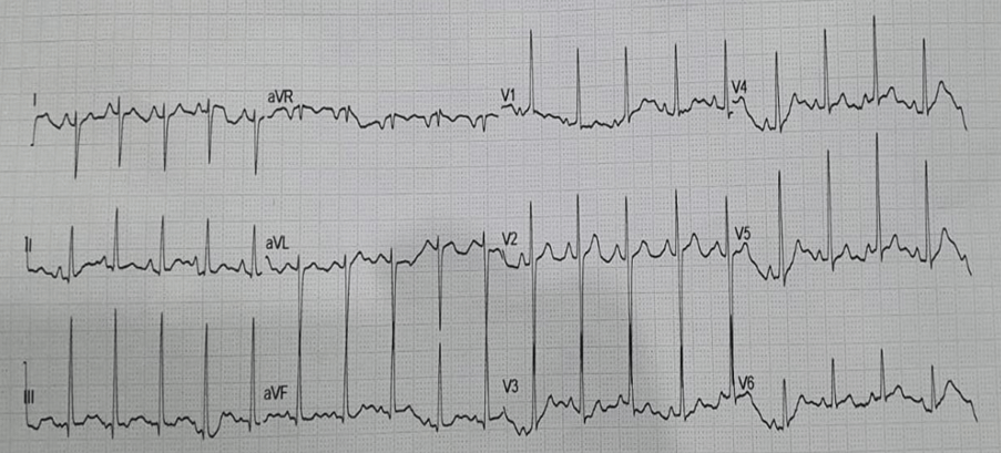
Findings:
- Right ventricular hypertrophy (RVH): Tall R in V1 (R:S ratio >1)
- RAE (P pulmonlae in lead II with prominent P wave >1.5 mm, most marked in V2)
- Right axis deviation (QRS axis) = +1100 (>+900)
- V2 sign: prominent R in lead V2 but less in amplitude compared to V1 and V3 …
- suggestive of large septal gap of VSD
- Sinus tachycardia
Comment
Suggestive of Tetralogy of Fallot
Chest X-ray PA-View
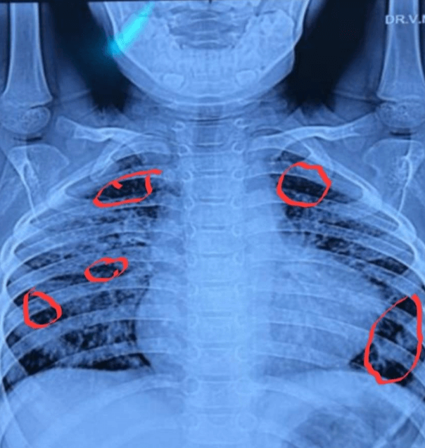
Findings
Boot-Shaped heart with upward turned cardiac apex due to the enlarged right Ventricle.
This CXR of TOF shows lungs major perfusion through collaterals and rest by whatever coming through pulmonary artery. In this CXR, it can be well appreciated that:
- Focal area of increased vascular marking
- Interspersed area of hyperlucent /oligaemic lung field (indicated by red circle mark on CXR)
- RT lung to left lung vascular marking asymmetry.
- Crisscrossing of vascular marking at many places
- The features described above is called LACY pattern of vascular distribution/marking
NB: Normal flow through pulmonary artery produces a uniform and bilateral almost symmetrically vascular markings from hilum to periphery – vessels almost parallel to but with tortuous course, as seen in most of CXR of TOF.
Discussion
Pulmonary hyperemia, defined as an abnormal increase in blood flow to the pulmonary vasculature, is not a typical feature of Tetralogy of Fallot (TOF), the associated condition of pulmonary stenosis as hallmark pathology reduces blood flow to the lungs resulting in an oligaemic lung field. However, 2 to 5% cases show pulmonary hyperemia in association with, making it a notable phenomenon in such select cases of TOF. Understanding its underlying
mechanisms is essential and crucial for managing such group of TOF and its complications.
Mechanism leading to pulmonary hyperemia in such cases of TOF Development of collaterals (aortic-bronchial and systemic-pulmonary collaterals)
Pulmonary hyperemia in TOF can be recognised by the radiological pattern.
This discussion highlights the significance of exploring hyperemia in the context of TOF, emphasing its role in understanding disease mechanisms, patients outcomes, andtherapeutic approaches.
Conclusion
Understanding the interplay between TOF and hyperemia is crucial for managing this condition. Assessing how the body attempts to compensate through hyperemic responses provides valuable insights into the severity of the defect and its physiological impact.
Furthermore, this knowledge is vital for optimizing surgical and medical interventions aimed at improving oxygen delivery and overall cardiac function.
References:
- Tetralogy of Fallot https://www.heart.org/en/health-topics/congenital-heart-defects/about-congenital-heart-defects/tetralogy-of-fallot
- Tetralogy of Fallot – Wikipedia https://en.wikipedia.org/wiki/Tetralogy_of_Fallot
- Tetralogy of Fallot Chest Xray, 14 April 2020 https://www.wikidoc.org/index.php/Tetralogy_of_fallot_chest_xray
- Tetralogy of Fallot – Radiopaedia https://radiopaedia.org/articles/tetralogy-of-fallot#:~:text=Chest%20radiographs%20may%20classically%20show,to%20decreased%20pulmonary%20arterial%20flow.
- Three steps approach for preoperative evaluation of tetralogy of Fallot patients: role of 128 MDCT Sherif Abd El Fattah Moustafa, et al, 04 Feb 2021 https://ejrnm.springeropen.com/articles/10.1186/s43055-021-00418-z
- Fallot Tetralogy https://www.sciencedirect.com/topics/nursing-and-health-professions/fallot-tetralogy
Discover CME INDIA:

- Explore CME INDIA Repository
- Examine CME INDIA Case Study
- Read History Today in Medicine
- Register for Future CMEs