

CME INDIA Presentation by Dr. Ashish Saxena MD, FRCP (Edin), FDIE, FRSSDI, FISH, MACP, Cardio-Diabeto-Metabolic Physician, Diabetes & Heart Centre, Ludhiana.
What the echo shows — and what the 2026 evidence tells us to do about it
Based on a lecture delivered at ECHO ASIA / ECHO INDIA 2026, Hall D, Leela Ambience, Gurugram (Delhi NCR), 9 October 2026.
| Key points The diabetic heart remodels years before the ejection fraction falls: LVH, diastolic dysfunction, reduced global longitudinal strain (GLS) and reduced left atrial (LA) reservoir strain mark Stage B — the treatable window. The ASE 2025 diastolic update uses e′, E/e′ and TR velocity, with LA reservoir strain ≤18% as the tie-breaker; diastolic stress echo unmasks HFpEF in the breathless patient with a normal resting study. The ESC 2026 heart failure guideline retires HFmrEF: an LVEF below 50% is now HFrEF, so the EF reported in the echo lab directly decides the drug list. Organ protection is phenotype-driven: SGLT2 inhibitors for heart failure and kidney; GLP-1 RA for atherosclerosis and weight; tirzepatide or semaglutide for obese HFpEF; finerenone for cardiorenal risk — combine when indications overlap. LDL-C goals are back (<55, <70, <100 mg/dL); combination lipid therapy should start early after an event. |
Introduction: the twin epidemic
Diabetes is no longer best understood as a disorder of glucose that occasionally injures the heart. It is a cardio-kidney-metabolic disease in which the myocardium is a principal target organ. The IDF Diabetes Atlas (2025) estimates 589 million adults with diabetes worldwide, projected to reach 853 million by 2050. India carries the second-largest burden — about 90 million adults by IDF estimates, and around 101 million with a further 136 million with prediabetes in ICMR-INDIAB. Cardiovascular mortality is two to four times higher in people with diabetes than in those without.
Three observations make this urgent for Indian practice. Heart failure — frequently heart failure with preserved ejection fraction (HFpEF) — is now a common first cardiovascular manifestation of type 2 diabetes. Chronic kidney disease (CKD), present in roughly 40% of people with type 2 diabetes, multiplies cardiovascular risk at every eGFR level. And the Asian-Indian phenotype develops diabetes, and its first myocardial infarction, about a decade earlier, with more visceral fat at a lower BMI. For the clinician holding the echo probe the lesson is simple: structural heart disease starts years before symptoms, and we must look for it.
This review follows one patient through three questions — why the diabetic heart fails, how to image it, and how to protect it — drawing on the 2025 ASE diastolic function update, the 2026 ESC heart failure guideline, the 2026 ADA Standards of Care and the cardiometabolic trials of the last two years.
| Case vignette — Mr K A 58-year-old IT professional from Pune with type 2 diabetes for 9 years (HbA1c 8.2% on metformin and glimepiride) and hypertension for 6 years (BP 152/94 mmHg on amlodipine 5 mg). BMI 29 kg/m², waist 102 cm, ex-smoker; father had a myocardial infarction at 56. He presented with three days of chest heaviness, now on one flight of stairs; heart rate 92/min with bibasal fine crackles. Work-up: ST depression V4–V6, raised troponin-I, NT-proBNP 1,820 pg/mL; LDL-C 142, triglycerides 248, ApoB 118 mg/dL; eGFR 62 mL/min/1.73 m², UACR 78 mg/g (CKD G2A2). Echo: LVEF 44%, mild LVH, grade 2 diastolic dysfunction. Angiography: 80% mid-LAD and 70% RCA stenoses; PCI to LAD. Diagnosis: NSTEMI with LVEF 44% — which under ESC 2026 is HFrEF. |
Why the diabetic heart fails differently?
The AHA cardiovascular-kidney-metabolic (CKM) construct places diabetes at the centre of a continuum that runs from adiposity through metabolic risk factors and subclinical heart or kidney disease to clinical cardiovascular disease. The 2026 ESC guideline now formally adopts heart failure stages A to D. Stage B (pre-heart failure) — a structural or functional cardiac abnormality, or raised natriuretic peptide or troponin, without symptoms — is precisely where echocardiography earns its place, because it identifies the patient who should receive organ-protective drugs now rather than after the first admission.
Four converging injuries drive the diabetic myocardium, and each leaves an echo footprint:
- Hyperglycaemia and advanced glycation end-products — collagen cross-linking, microvascular dysfunction and oxidative stress produce a stiff ventricle: low e′, high E/e′.
- Insulin resistance and lipotoxicity — ectopic and epicardial fat with mitochondrial energetic failure: thicker epicardial adipose tissue, reduced GLS.
- RAAS and sympathetic over-activity — sodium retention, fibrosis, hypertrophy and arrhythmia: LVH, concentric remodelling, LA enlargement.
- Metaflammation — IL-6, TNF-α, NLRP3 and epicardial adipose signalling: coronary microvascular disease and the HFpEF phenotype.
Imaging the diabetic heart
The echo cascade
Diabetic cardiomyopathy unfolds as a recognisable sequence: concentric remodelling or LVH, then diastolic dysfunction, then subclinical systolic dysfunction (reduced GLS with a normal EF), then HFpEF, and finally HFrEF (Figure 1). The first three steps constitute Stage B. The disease is not a single phenotype — the restrictive, HFpEF-type pattern is commonest in type 2 diabetes, while a dilated, HFrEF-type picture usually reflects coexisting coronary disease. Ischaemia, hypertension, valve disease and infiltrative disease must be excluded before the label “diabetic cardiomyopathy” is applied. Strain and LA function are the most sensitive early signals; the ejection fraction is the last to fall.
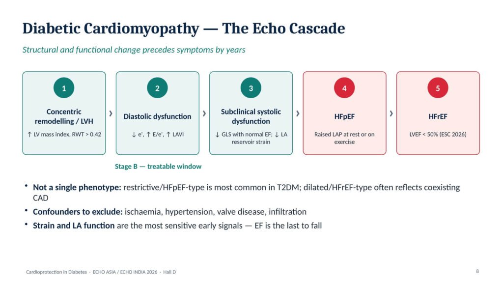
Figure 1. The echo cascade of diabetic cardiomyopathy. Concentric remodelling, diastolic dysfunction and subclinical systolic dysfunction define Stage B — the treatable window before HFpEF or HFrEF.
A standard protocol for every patient with diabetes
A focused diabetic-heart protocol (Figure 2) covers six domains: structure (LV mass index, relative wall thickness), systolic function (biplane LVEF and GLS), diastolic function (septal and lateral e′, average E/e′, E/A), the left atrium (LA volume index and LA reservoir strain), pressures (TR velocity / PASP), and epicardial fat and right-ventricular function, which should be reported and tracked over time. GLS thresholds are vendor-dependent, so each laboratory should use its own normal range.
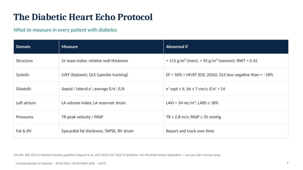
Figure 2. The diabetic heart echo protocol, with abnormal cut-offs from the ASE 2025 diastolic function update and the ESC 2026 heart failure guideline.
ASE 2025: diastolic function, updated
The American Society of Echocardiography update (Nagueh et al., July 2025) replaces the 2016 ASE/EACVI document and, importantly, brings LA strain into the algorithm [3]. In sinus rhythm, three main variables are assessed: reduced e′ (septal ≤6, lateral ≤7, or average ≤6.5 cm/s), average E/e′ >14, and TR velocity ≥2.8 m/s or PASP ≥35 mmHg. If none is abnormal, LA pressure is normal; if all three are abnormal, it is raised. When the findings are discordant, supplementary markers decide — LA reservoir strain (LARS) ≤18%, pulmonary venous S/D ≤0.67, LAVI >34 mL/m² or IVRT ≤70 ms (Figure 3). Grading is then straightforward: grade 1 with normal LA pressure and E/A ≤0.8; grade 2 with raised LA pressure and E/A <2; grade 3 with raised pressure and E/A ≥2. Age-specific e′ cut-offs reduce over-diagnosis in the elderly.
Up to half of patients with HFpEF have normal filling pressures at rest. For the breathless diabetic with a “normal” resting echo, diastolic stress echocardiography is the logical next step: an average E/e′ ≥14 (septal ≥15) with peak TR velocity >3.2 m/s is abnormal, while E/e′ <10 with TR <2.8 m/s is normal.
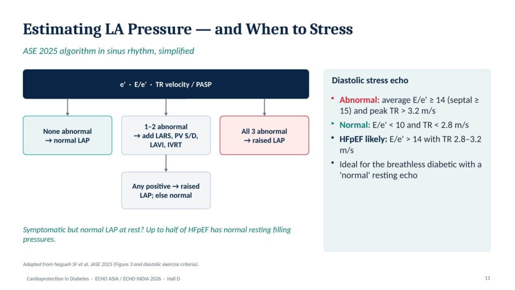
Figure 3. Estimating LA pressure in sinus rhythm (ASE 2025, simplified), and criteria for diastolic stress echocardiography. Adapted from Nagueh et al., JASE 2025.
Strain: the early systolic and atrial signals
Diabetic hearts lose longitudinal function while the EF is still normal. A meta-analysis of 41 studies in asymptomatic adults (6,668 with diabetes, 7,218 controls) found GLS roughly 2 percentage points lower in diabetes (mean difference −1.96%), with the largest effect in LA reservoir strain (mean difference −8.4) [4]. Higher BMI was associated with worse GLS and higher HbA1c with worse RV strain (Figure 4); in longitudinal cohorts, reduced GLS predicts incident heart failure and death. Heterogeneity between studies was high, which reinforces the need for consistent vendor and software when strain is used serially.
LA reservoir strain — atrial filling during ventricular systole — falls first as the ventricle stiffens. It serves as the ASE 2025 tie-breaker for indeterminate e′/E/e′/TR combinations and, usefully, improves as filling pressures fall on treatment.
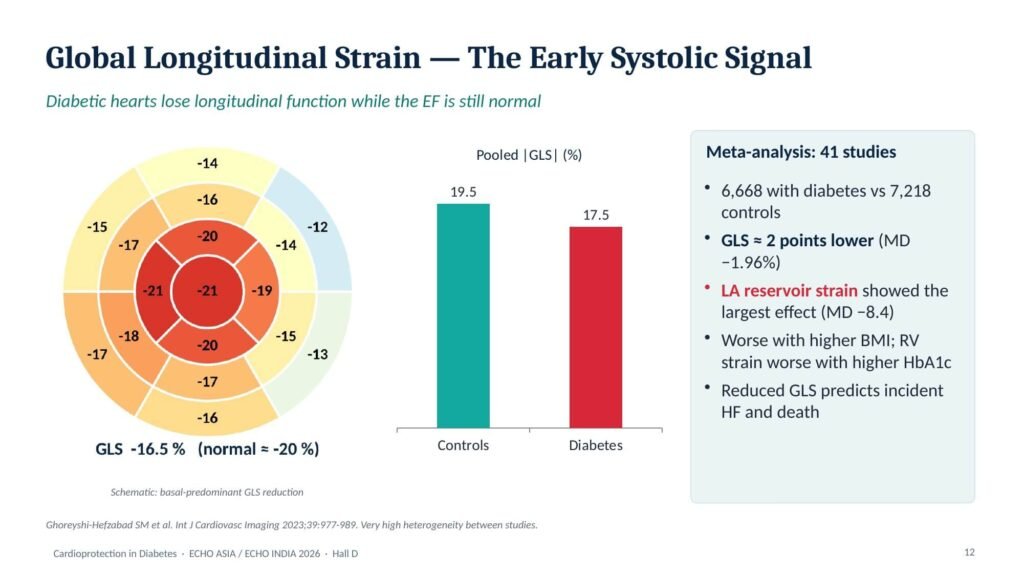
Figure 4. Global longitudinal strain in diabetes: basal-predominant GLS reduction (schematic) and pooled |GLS| in controls versus diabetes. Data from Ghoreyshi-Hefzabad et al., 2023.
Whom to image?
A practical pathway is “biomarker first, echo second, treatment immediately” (Figure 5). The ADA Standards support annual NT-proBNP or high-sensitivity troponin in adults with diabetes [1]; an NT-proBNP ≥125 pg/mL or raised hs-troponin should prompt transthoracic echocardiography, and any Stage B abnormality should trigger organ protection. Image directly, without waiting for biomarkers, in patients with symptoms, atrial fibrillation, an abnormal ECG or murmur; in long-standing diabetes with hypertension or CKD; and whenever the EF will change the drug list. Repeat imaging at 3–6 months after therapy is optimised.
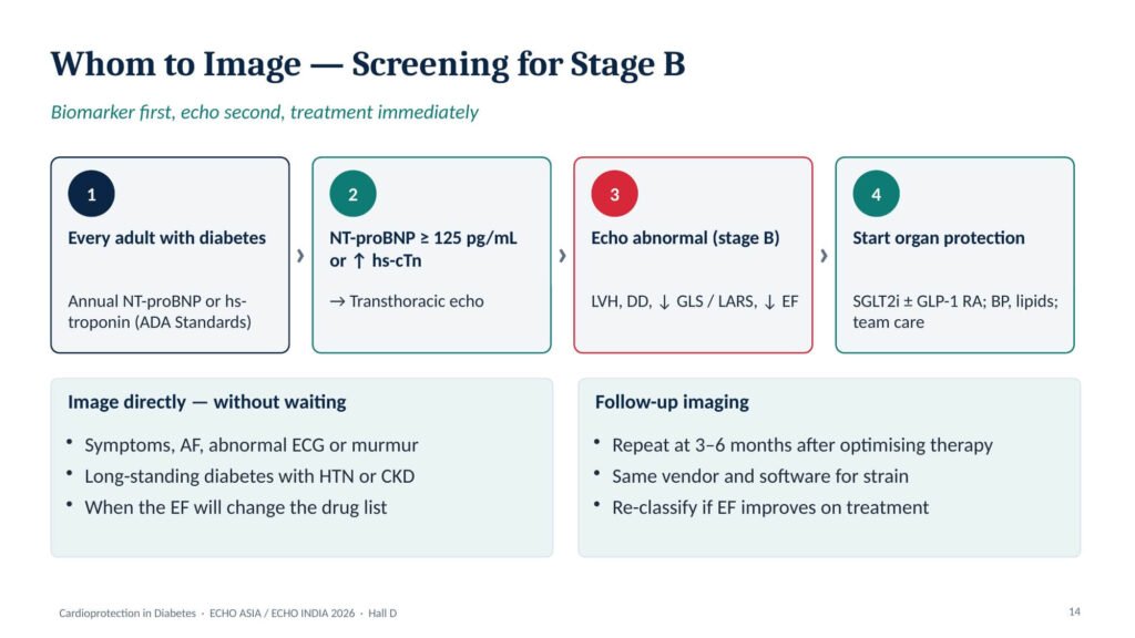
Figure 5. Screening for Stage B heart failure in diabetes: annual biomarker testing, echocardiography if abnormal, and immediate organ protection.
Seeing cardioprotection
These drugs remodel the diabetic heart, and imaging can measure it. In EMPA-HEART, empagliflozin regressed LV mass index on CMR at six months in people with type 2 diabetes and coronary disease; EMPA-TROPISM showed reduced LV volumes and mass with a rise in LVEF in non-diabetic HFrEF. The STEP-HFpEF echo sub-study showed favourable LA remodelling with semaglutide 2.4 mg, and the SUMMIT CMR sub-study showed reductions in LV mass and paracardiac adipose tissue with tirzepatide.
ESC 2026: a simpler heart failure map
The 2026 ESC guideline, presented in Munich in August 2026, eliminates heart failure with mildly reduced EF [2]. HFrEF is now an LVEF below 50%, and HFpEF an LVEF of 50% or above with objective evidence of cardiac abnormality; “decompensated” heart failure replaces “acute” heart failure. ACEi/ARNI, beta-blocker, MRA and SGLT2 inhibitor form the foundational therapy for HFrEF; MRA and SGLT2 inhibitors are foundational across the EF spectrum; and semaglutide or tirzepatide is recommended (Class IIa) in HFpEF with BMI ≥30 kg/m² (Figure 6).
For the echo laboratory the consequence is direct: the EF you report decides the drug list. Mr K’s LVEF of 44% — formerly HFmrEF — is now HFrEF with Class I foundational therapy. Measurement error around 50% matters, so biplane Simpson’s (or 3D) should be used and repeated after therapy.
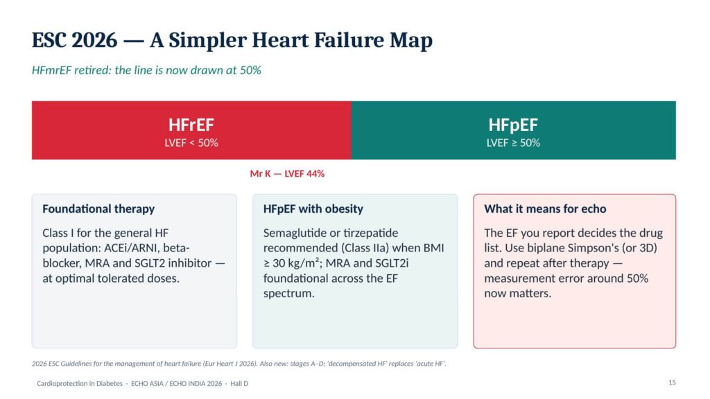
Figure 6. The ESC 2026 heart failure map: HFmrEF is retired and the HFrEF/HFpEF boundary is drawn at an LVEF of 50%.
Protecting the diabetic heart
The 2025–26 guideline wave
Six documents point in one direction — organ protection first. The ADA Standards 2026 make cardiorenal protection co-equal with glucose lowering, endorse SBP <120 mmHg if tolerated in high-risk patients, and allow an SGLT2 inhibitor and finerenone to be started together [1]. The 2026 ACC/AHA multisociety dyslipidaemia guideline restores LDL-C goals and adopts the PREVENT equations [5]. The ESC/EAS 2025 focused update advises statin plus ezetimibe upfront after ACS. The AHA/ACC 2025 documents set a BP goal of <130/80 mmHg and prefer ticagrelor or prasugrel over clopidogrel after ACS. The ESC 2026 heart failure guideline is described above, and the ESC 2026 CVD and CKD guidance asks for eGFR and albuminuria in every cardiovascular patient. Together they define four pillars: metabolic, haemodynamic, lipid and antithrombotic.
SGLT2 inhibitors: heart failure is the signal
Across the class, SGLT2 inhibitors reduce heart failure hospitalisation by about 30%. Cardiovascular outcome trials (EMPA-REG, CANVAS, DECLARE, VERTIS) showed 27–35% fewer HF admissions; in HFrEF, DAPA-HF and EMPEROR-Reduced gave hazard ratios of 0.74–0.75 for CV death or worsening HF, and in HFpEF, EMPEROR-Preserved and DELIVER gave 0.79–0.82. In CKD (CREDENCE, DAPA-CKD, EMPA-KIDNEY), kidney risk fell by 28–39% down to an eGFR of 20. After myocardial infarction (EMPACT-MI) the benefit was fewer HF admissions rather than fewer MACE.
GLP-1 receptor agonists: atherosclerosis, kidney, weight
A meta-analysis of eight cardiovascular outcome trials showed GLP-1 RAs reduce MACE by 14%, stroke by 17% and the kidney composite by 21%. SELECT extended a 20% MACE reduction to people with obesity and cardiovascular disease without diabetes; SOUL established the first oral GLP-1 RA with cardiovascular benefit (MACE −14%); and FLOW, in type 2 diabetes with CKD, reduced the kidney composite by 24% and CV death by 29% [7].
Obese HFpEF: the incretin breakthrough
The STEP-HFpEF programme (529 patients without and 616 with diabetes, BMI ≥30) showed improved symptoms (KCCQ-CSS +7.8 points in the non-diabetic trial), weight loss of about 10%, a 20 m gain in six-minute walk distance, lower hs-CRP and NT-proBNP, and favourable LA remodelling with semaglutide 2.4 mg [6]. SUMMIT (731 patients, mean BMI 38) was the first incretin trial to show an HF-event benefit: tirzepatide reduced CV death or worsening HF (HR 0.62), improved KCCQ-CSS by 6.9 points, produced 13.9% versus 2.2% weight loss, and reduced LV mass and paracardiac fat on CMR [8]. Both ADA 2026 and ESC 2026 now support these agents in obese HFpEF.Tirzepatide and finerenone: completing the toolkit
In SURPASS-CVOT (13,165 people with type 2 diabetes and ASCVD, about four years), tirzepatide was non-inferior to dulaglutide for 3-point MACE (HR 0.92, 95% CI 0.83–1.01) without proving superiority, while producing greater reductions in HbA1c, weight, systolic BP and ApoB [9]. The non-steroidal MRA finerenone reduced the kidney composite by 18% in FIDELIO-DKD, the CV composite by 13% (with less new HF) in FIGARO-DKD, and CV death or worsening HF by 16% in FINEARTS-HF (EF ≥40%) [10]. In CONFIDENCE, combining finerenone with empagliflozin lowered UACR by 52% compared with about 30% for either alone. Check potassium at four weeks; SGLT2 inhibitor co-therapy mitigates hyperkalaemia.
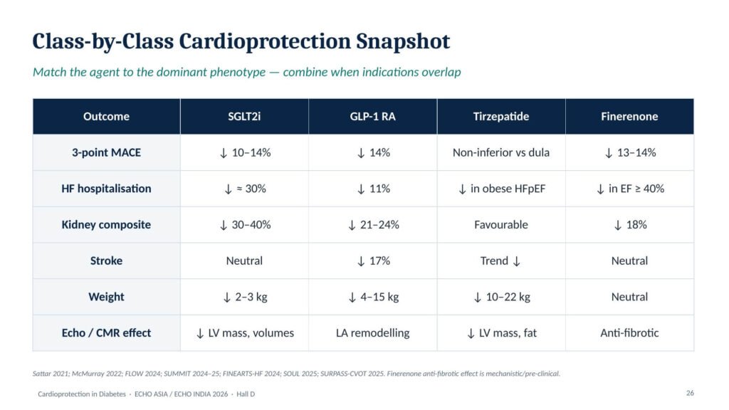
Figure 7. Class-by-class cardioprotection snapshot: SGLT2 inhibitors, GLP-1 RA, tirzepatide and finerenone compared across MACE, HF, kidney, stroke, weight and imaging effects.
Lipids, blood pressure and antithrombotic therapy
LDL-C goals are back: <55 mg/dL for very-high-risk ASCVD, <70 mg/dL for high risk and <100 mg/dL for borderline or intermediate risk [5]. In diabetes, primary-prevention lipid-lowering therapy is indicated regardless of baseline LDL-C; Lp(a) should be measured once in adulthood, ApoB when triglycerides are high, and coronary calcium when risk is uncertain. Therapy escalates from a high-intensity statin to ezetimibe (upfront if very high risk), bempedoic acid, and a PCSK9 monoclonal antibody or inclisiran if LDL-C remains ≥55 mg/dL at 4–8 weeks.
Blood pressure should be below 130/80 mmHg, with SBP <120 mmHg if tolerated in high cardiovascular or kidney risk. ACEi or ARB is first-line, particularly with albuminuria, followed by a calcium-channel blocker or chlorthalidone, and ARNI in HFrEF; SGLT2 inhibitors add a 3–5 mmHg systolic reduction. Routine aspirin is not recommended for primary prevention (ASCEND, ASPREE). After ACS, ticagrelor or prasugrel is preferred over clopidogrel, with ticagrelor monotherapy after one month as an option; in chronic coronary or peripheral disease with low bleeding risk, rivaroxaban 2.5 mg twice daily plus aspirin may be added. Diabetes increases bleeding risk on dual antiplatelet therapy, so individualise.
From echo phenotype to prescription
The bedside algorithm (Figure 8) matches therapy to the dominant phenotype. HFrEF (LVEF <50%): ARNI/ACEi, beta-blocker, MRA and SGLT2 inhibitor, started early and in hospital once stable. HFpEF (LVEF ≥50%): SGLT2 inhibitor and MRA/finerenone, adding semaglutide or tirzepatide if BMI ≥30. Stage B: SGLT2 inhibitor ± GLP-1 RA, BP <130/80 and a repeat echo at 3–6 months. Diabetes with ASCVD: GLP-1 RA (or tirzepatide) ± SGLT2 inhibitor, statin plus ezetimibe to LDL <55. Diabetes with CKD (UACR ≥30): RAS inhibitor, SGLT2 inhibitor and finerenone (which may start together), then semaglutide. For every patient: metformin and lifestyle, BP and LDL to goal, annual UACR, eGFR and NT-proBNP, smoking cessation, OSA screening and vaccination.
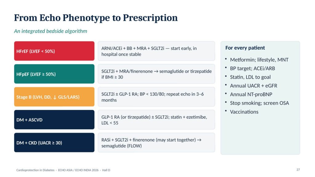
Figure 8. An integrated bedside algorithm from echo phenotype to prescription, with measures that apply to every patient with diabetes.
Back to Mr K
Mr K — NSTEMI, HFrEF (EF 44%), CKD G2A2, obesity and LDL-C 142 mg/dL — leaves hospital on aspirin 75 mg plus ticagrelor 90 mg twice daily (with ticagrelor alone considered after one month); rosuvastatin 40 mg with ezetimibe 10 mg from day one, targeting LDL-C <55 mg/dL; ramipril with planned switch to ARNI, bisoprolol, empagliflozin 10 mg and an MRA (finerenone if potassium <5.0 mmol/L); and semaglutide titrated from 0.25 to 1 mg weekly for ASCVD, weight and glycaemia. Glimepiride is stopped, metformin continued, and SBP targeted below 130 mmHg (below 120 if tolerated). At three months his echo will re-measure biplane LVEF (re-classifying him if it improves), GLS and LA reservoir strain, LV mass index, LAVI, E/e′ and TR velocity, paired with NT-proBNP, potassium, eGFR and UACR.
| Take-home messages Look early. The diabetic heart changes years before the EF: LVH, diastolic dysfunction, reduced GLS and reduced LA strain. Use ASE 2025. e′, E/e′ and TR first, then LA strain as the tie-breaker; stress echo for the breathless patient with a normal resting study. Report EF carefully. Under ESC 2026, an LVEF <50% is HFrEF — the number decides the prescription. Screen for Stage B. Annual NT-proBNP or hs-troponin, then echo, then treat immediately. Phenotype-driven therapy. SGLT2i for HF and kidney; GLP-1 RA for MACE and weight; finerenone for cardiorenal risk — combine. Lower LDL, earlier. <55 mg/dL after an event, with combination therapy upfront. |
Echo figures reproduced from the lecture slides are schematic illustrations, not patient images.
References:
- American Diabetes Association Professional Practice Committee. Standards of Care in Diabetes—2026. Diabetes Care 2026;49(Suppl 1).
- Køber L, Adamo M, Ruwald A-C, et al. 2026 ESC Guidelines for the management of heart failure. Eur Heart J 2026. doi:10.1093/eurheartj/ehag100.
- Nagueh SF, Sanborn DY, Oh JK, et al. Recommendations for the evaluation of left ventricular diastolic function by echocardiography and for heart failure with preserved ejection fraction diagnosis: an update from the American Society of Echocardiography. J Am Soc Echocardiogr 2025;38:537–69.
- Ghoreyshi-Hefzabad SM, Jeyaprakash P, Vo HQ, et al. Subclinical systolic dysfunction detected by 2D speckle tracking echocardiography in adults with diabetes mellitus: systematic review and meta-analysis of 6668 individuals with diabetes mellitus and 7218 controls. Int J Cardiovasc Imaging 2023;39:977–89.
- Blumenthal RS, Morris PB, Gaudino M, et al. 2026 ACC/AHA/Multisociety guideline on the management of dyslipidemia. J Am Coll Cardiol 2026. doi:10.1016/j.jacc.2025.11.016.
- Kosiborod MN, Petrie MC, Borlaug BA, et al. Semaglutide in patients with obesity-related heart failure and type 2 diabetes (STEP-HFpEF DM). N Engl J Med 2024;390:1394–407.
- Perkovic V, Tuttle KR, Rossing P, et al. Effects of semaglutide on chronic kidney disease in patients with type 2 diabetes (FLOW). N Engl J Med 2024;391:109–21.
- Packer M, Zile MR, Kramer CM, et al. Tirzepatide for heart failure with preserved ejection fraction and obesity (SUMMIT). N Engl J Med 2025;392:427–37.
- Nicholls SJ, Pavo I, Bhatt DL, et al. Cardiovascular outcomes with tirzepatide versus dulaglutide in type 2 diabetes (SURPASS-CVOT). N Engl J Med 2025.
- Solomon SD, McMurray JJV, Vaduganathan M, et al. Finerenone in heart failure with mildly reduced or preserved ejection fraction (FINEARTS-HF). N Engl J Med 2024;391:1475–85.

Discover CME INDIA

- Explore CME INDIA Repository
- Examine CME INDIA Case Study
- Read History Today in Medicine
- Register for Future CMEs