
CME INDIA Case Presentation by Dibyendu Dey, BALCO Medical Centre, Naya Raipur, Chhattisgarh, India.
CME INDIA Case Study
Think beyond the pain crisis — and check the reticulocyte count.
| ABSTRACT Background: Transient aplastic crisis (TAC) is an acute, potentially fatal interruption of erythropoiesis in patients with chronic haemolytic anaemias, most commonly caused by human parvovirus B19. In sickle cell disease (SCD) the shortened red-cell lifespan converts a brief marrow pause into profound anaemia. Case presentation: A 21-year-old man with homozygous sickle cell anaemia (HbSS), previously transfusion-independent with only one or two lifetime pain crises, developed weakness after two days of mild fever. Haemoglobin (Hb) was 4.1 g/dL and, despite two units of packed red cells given elsewhere, 2.8 g/dL on arrival. There was no pain, jaundice or bleeding. The blood picture showed normocytic normochromic anaemia with preserved leucocyte and platelet lines; liver and renal function and lactate dehydrogenase (LDH) were normal, and the spleen was small (~5 cm). The case discussion identified parvovirus B19–associated aplastic crisis. Conclusion: Fever followed by an abrupt Hb fall in SCD, without haemolytic markers or splenomegaly, should prompt an immediate absolute reticulocyte count and parvovirus B19 testing. A further Hb fall after transfusion is not explained by aplasia alone and warrants separate evaluation for haemolytic transfusion reaction or hyperhaemolysis. Keywords: sickle cell disease; parvovirus B19; transient aplastic crisis; reticulocytopenia; hyperhaemolysis; India |
Introduction
Sickle cell disease is a major public-health problem in India, concentrated in tribal and other communities of central, western and southern India, including Chhattisgarh, Madhya Pradesh, Maharashtra, Gujarat and Odisha.1 The clinical course of HbSS in India is notably heterogeneous; co-inheritance of α-thalassaemia and high fetal haemoglobin (HbF) associated with the Arab-Indian haplotype contribute to a milder phenotype in many patients.1,2 A mild baseline course, however, does not protect against acute life-threatening complications.
Acute anaemic events in SCD — an Hb fall of ≥2 g/dL below steady state — have three principal mechanisms: failure of red-cell production (aplastic crisis), pooling of blood in an organ (splenic or hepatic sequestration), and accelerated destruction (hyperhaemolysis, including delayed haemolytic transfusion reactions).2,3 The association of aplastic crisis with parvovirus B19 was established in 1981, and the virus is now recognised as responsible for the large majority of such episodes.4–6 We describe a young adult with a previously benign course who presented with profound anaemia after a brief febrile illness.
Case Presentation
History
A 21-year-old man with known homozygous sickle cell anaemia had been largely asymptomatic, with only one or two vaso-occlusive pain episodes in his lifetime and no prior blood transfusions. He was taking folic acid intermittently and was not on hydroxyurea. He presented with progressive weakness following two days of mild fever. He denied bone or chest pain, jaundice, dark urine, haematemesis, melaena or other bleeding.
At a local facility, Hb was 4.1 g/dL and two units of packed red cells were transfused. On arrival at our centre, repeat Hb was 2.8 g/dL. The exact interval between transfusion and the repeat sample was not available.
Examination
He was markedly pale. There was no icterus, lymphadenopathy, bleeding manifestation or palpable hepatosplenomegaly. Systemic examination was otherwise unremarkable.
Investigations
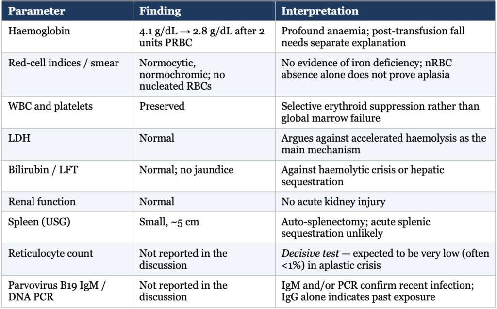
Key results are summarised in Table 1. The peripheral smear showed normocytic, normochromic anaemia with preserved white cell and platelet lines and no nucleated red cells. Liver and renal function tests and LDH were within normal limits. Ultrasonography showed a small spleen measuring approximately 5 cm, consistent with auto-splenectomy.
Table 1. Clinical and laboratory findings and their interpretation
Diagnosis
The clinical case discussion on the CME India platform identified the diagnosis as transient aplastic crisis associated with parvovirus B19. The pattern — brief febrile prodrome, abrupt severe anaemia, preserved leucocytes and platelets, normal haemolytic markers and a small spleen — is characteristic. Confirmation rests on a markedly reduced absolute reticulocyte count with parvovirus B19 IgM and/or DNA PCR; the specific test and result used were not provided in the discussion.
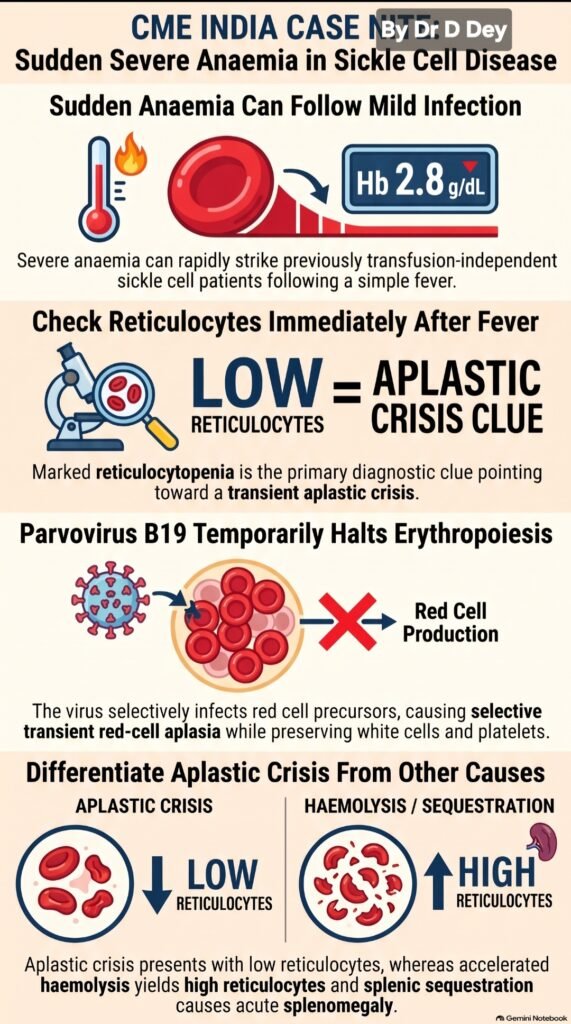
Figure 1. Visual abstract of the case (CME India case note infographic, prepared by Dr D. Dey). Key messages: sudden anaemia can follow a mild infection; check reticulocytes immediately; parvovirus B19 halts erythropoiesis; differentiate aplastic crisis (low reticulocytes) from haemolysis or sequestration (high reticulocytes).
Discussion
Pathophysiology
Parvovirus B19 is a small single-stranded DNA virus with marked tropism for erythroid progenitor cells. It enters cells through blood group P antigen (globoside) together with co-receptors, replicates in BFU-E and CFU-E, and causes their lysis, arresting red-cell production for roughly one week.7,8 In haematologically normal individuals, whose red cells survive about 120 days, this pause produces only a trivial Hb fall. In HbSS, red-cell survival is reduced to approximately 10–20 days, and the same pause can halve the Hb concentration within days (Figure 2).2,7
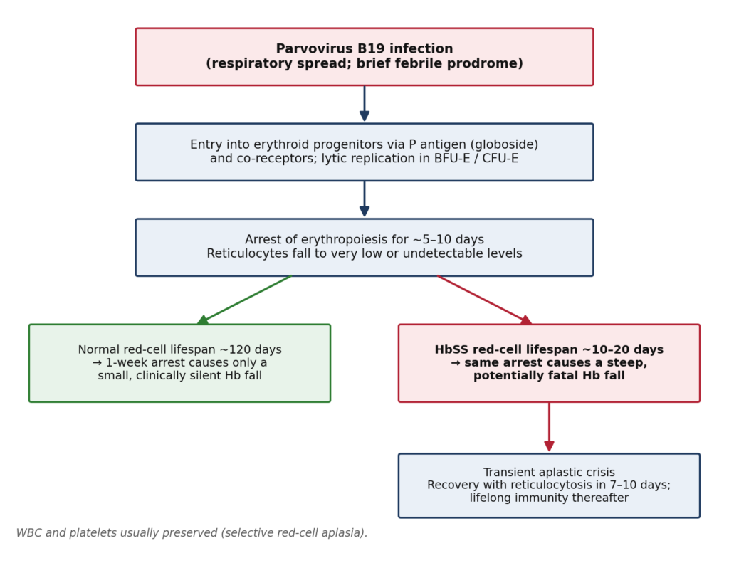
Figure 2. Pathophysiology of parvovirus B19–associated transient aplastic crisis. The same transient erythroid arrest is clinically silent with normal red-cell survival but life-threatening when red-cell lifespan is shortened, as in sickle cell disease.
Clinical course and diagnosis
Typical TAC begins with fever, malaise, headache or gastrointestinal symptoms, followed by fatigue, pallor and breathlessness; the classic “slapped cheek” rash is uncommon in this setting.5,6 The defining laboratory feature is reticulocytopenia — frequently below 1% — in a patient whose steady state normally shows brisk reticulocytosis (Figure 3). Neutropenia and thrombocytopenia may occasionally accompany the episode but are usually mild.6 Recovery is heralded by a surge of reticulocytes and nucleated red cells 7–10 days after onset, after which immunity is generally lifelong.5,7
Serology and molecular testing establish the aetiology. B19 IgM appears within about 10–12 days of infection; DNA PCR detects viraemia and is particularly useful early, when IgM may still be negative. A positive IgG alone indicates past infection.7 Bone-marrow examination, when performed, shows absent or markedly reduced erythroid precursors with occasional giant pronormoblasts, but is rarely necessary.
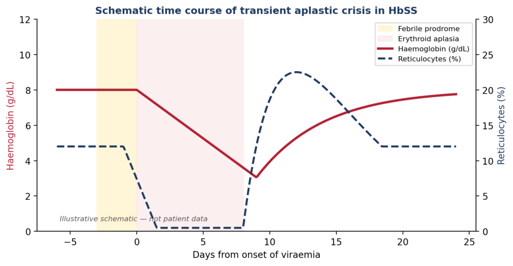
Figure 3. Schematic time course of transient aplastic crisis in HbSS: reticulocytes fall shortly after viraemia, haemoglobin declines steadily during the aplastic phase, and recovery is marked by a reticulocyte surge. Illustrative only; not patient data.
Differential diagnosis
The reticulocyte count, spleen size and haemolytic markers together discriminate between the major mechanisms of acute anaemia in SCD (Table 2; Figure 4).2,3,9,10 In adults with HbSS, auto-splenectomy makes splenic sequestration uncommon, although hepatic sequestration should still be considered.
Table 2. Distinguishing acute anaemic events in sickle cell disease
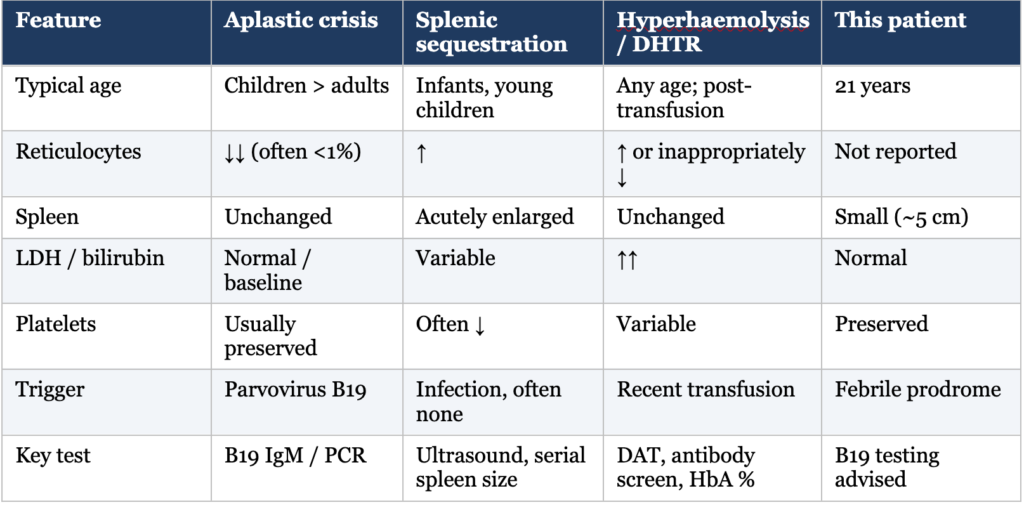
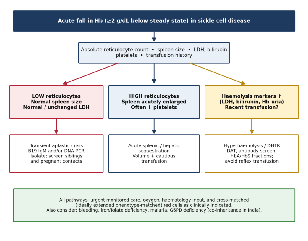
Figure 4. Practical diagnostic approach to an acute fall in haemoglobin in sickle cell disease. DHTR, delayed haemolytic transfusion reaction; DAT, direct antiglobulin test.
The post-transfusion haemoglobin paradox
Two units of packed red cells would be expected to raise Hb by roughly 2 g/dL in an adult. The subsequent value of 2.8 g/dL therefore implies a fall of approximately 3 g/dL from the anticipated post-transfusion level. Aplasia alone halts production but cannot account for such rapid loss of transfused cells over a short interval. Possible explanations include a laboratory or sampling error (for example, a sample drawn from an infusion limb), incomplete transfusion, concealed bleeding, and — most importantly — an acute or delayed haemolytic transfusion reaction or hyperhaemolysis syndrome, in which both donor and autologous red cells are destroyed.9,10
The Hb should be confirmed on a fresh sample, and the transfusion timeline and any immediate post-transfusion value reviewed. If the fall is genuine, evaluation should include serial LDH and bilirubin, urine for haemoglobin, a direct antiglobulin test, a red-cell antibody screen, and HbA/HbS quantification to assess survival of donor cells. Normal LDH makes significant haemolysis less likely, but a negative DAT does not exclude hyperhaemolysis.10 Recognising this possibility matters because reflexive further transfusion may worsen hyperhaemolysis.
Management
Treatment of TAC is supportive. Patients with symptomatic or profound anaemia require prompt simple transfusion, ideally with ABO-, Rh (C, c, E, e)- and Kell-matched red cells, aiming to restore approximately steady-state Hb and avoiding over-transfusion (generally above 10 g/dL) because of hyperviscosity.3,11 Haemodynamic monitoring, oxygen and close observation for acute chest syndrome, stroke and other complications are essential; neurological and pulmonary events have been reported during parvovirus infection in SCD.6 Daily reticulocyte counts track recovery. Intravenous immunoglobulin is reserved for persistent B19 infection in immunocompromised hosts and is not routinely needed in TAC.7
Infection-control measures are important. Patients are viraemic and infectious during the aplastic phase; droplet precautions are advised, and pregnant staff and contacts with haemolytic disorders or immunodeficiency should avoid exposure. Siblings with SCD should be monitored for pallor and fatigue over the following weeks, since household clustering is common.4,5
Relevance to Indian practice
With the launch of the National Sickle Cell Anaemia Elimination Mission in 2023, screening and long-term care of SCD in India are expanding, and more adults with milder phenotypes will be known to physicians outside specialist centres. Internists in endemic regions should keep TAC in mind whenever a febrile illness is followed by sudden severe pallor, and should simultaneously exclude malaria, co-inherited G6PD deficiency and nutritional deficiencies. A reticulocyte count is inexpensive, widely available and diagnostically decisive.
CME INDIA Clinical Pearls
- A mild past course does not exclude a severe acute complication. Even a transfusion-independent patient with SCD can develop profound anaemia after an infection.
- Fever followed by sudden anaemia should trigger a reticulocyte count. Use the absolute reticulocyte count; marked reticulocytopenia is the key clue to aplastic crisis.
- Parvovirus B19 temporarily switches off red-cell production. In SCD the shortened red-cell lifespan makes this pause potentially life-threatening.
- Preserved white cells and platelets support selective erythroid suppression. “Aplastic crisis” here means transient red-cell aplasia, not pancytopenic aplastic anaemia.
- Distinguish the mechanisms of sudden anaemia. Aplasia: low reticulocytes. Haemolysis: raised LDH/bilirubin with reticulocytosis. Sequestration: acute organ enlargement, often thrombocytopenia.
- Establish the viral aetiology. B19 IgM and/or DNA PCR support recent infection; IgG alone indicates prior exposure.
- A falling Hb after transfusion deserves independent evaluation. Confirm the value and timeline and assess for haemolytic transfusion reaction or hyperhaemolysis.
- Profound anaemia requires urgent monitored care. Stabilise and involve haematology while the diagnostic work-up proceeds.
| TAKE-HOME MESSAGE: In sickle cell disease, sudden anaemia after fever demands a reticulocyte count. A profoundly low count points towards transient aplastic crisis — most commonly due to parvovirus B19. A further fall in haemoglobin after transfusion should never be attributed to aplasia without excluding haemolysis. |
References:
- Colah RB, Mukherjee MB, Martin S, Ghosh K. Sickle cell disease in tribal populations in India. Indian J Med Res. 2015;141(5):509-515.
- Kato GJ, Piel FB, Reid CD, et al. Sickle cell disease. Nat Rev Dis Primers. 2018;4:18010.
- Yawn BP, Buchanan GR, Afenyi-Annan AN, et al. Management of sickle cell disease: summary of the 2014 evidence-based report by expert panel members. JAMA. 2014;312(10):1033-1048.
- Pattison JR, Jones SE, Hodgson J, et al. Parvovirus infections and hypoplastic crisis in sickle-cell anaemia. Lancet. 1981;1(8221):664-665.
- Serjeant GR, Topley JM, Mason K, et al. Outbreak of aplastic crises in sickle cell anaemia associated with parvovirus-like agent. Lancet. 1981;2(8247):595-597.
- Smith-Whitley K, Zhao H, Hodinka RL, et al. Epidemiology of human parvovirus B19 in children with sickle cell disease. Blood. 2004;103(2):422-427.
- Young NS, Brown KE. Parvovirus B19. N Engl J Med. 2004;350(6):586-597.
- Brown KE, Anderson SM, Young NS. Erythrocyte P antigen: cellular receptor for B19 parvovirus. Science. 1993;262(5130):114-117.
- Emond AM, Collis R, Darvill D, et al. Acute splenic sequestration in homozygous sickle cell disease: natural history and management. J Pediatr. 1985;107(2):201-206.
- Danaee A, Inusa B, Howard J, Robinson S. Hyperhemolysis in patients with hemoglobinopathies: a single-center experience and review of the literature. Transfus Med Rev. 2015;29(4):220-230.
- Chou ST, Alsawas M, Fasano RM, et al. American Society of Hematology 2020 guidelines for sickle cell disease: transfusion support. Blood Adv. 2020;4(2):327-355.
- Goldstein AR, Anderson MJ, Serjeant GR. Parvovirus associated aplastic crisis in homozygous sickle cell disease. Arch Dis Child. 1987;62(6):585-588.
Discover CME INDIA:

- Explore CME INDIA Repository
- Examine CME INDIA Case Study
- Read History Today in Medicine
- Register for Future CMEs